Summary
Biomedical and biological materials inside the human body are complex structures consisting of multiple components. The mechanical interactions between different components in these structures governs their critical behaviors and performance. However, the underlying mechanics controlling these processes are not fully understood. Our research contributes to this understanding by focusing on the design, fabrication and characterization of these medical related materials and structures. It combines experimental measurements with analytical and computational models that enable the prediction of mechanical behaviors at the nano-, micro- and macro- scales. Interdisciplinary in essence, our research has both fundamental and applied aspects that are woven together through mechanics, materials and bioengineering.
Advanced Biomaterials for Endovascular Embolization
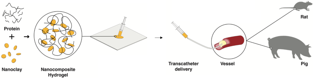
Minimally invasive transcatheter embolization is a common nonsurgical procedure in interventional radiology used for the deliberate occlusion of blood vessels for the treatment of diseased or injured vasculature. A wide variety of embolic agents including metallic coils, calibrated microspheres, and liquids are available for clinical practice. Additionally, advances in biomaterials, such as shape‐memory foams, biodegradable polymers, and in situ gelling solutions have led to the development of novel preclinical embolic agents. We aim to develop next-generation embolic agents and emerging technologies for endovascular embolization.
Relevant publication: Adv. Mater. (2019), Prog. Biomed. Eng. (2020), Adv. Mater. (2020), Adv. Mater. (2020), Adv. Sci. (2020)
Tumor-specific Nanoparticles
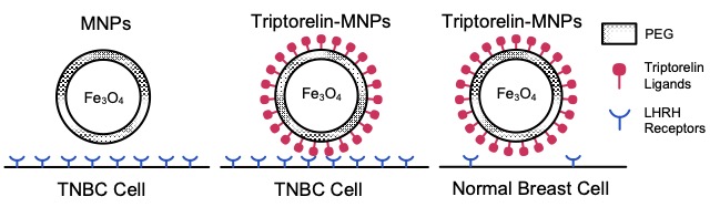
Targeted therapy is an emerging technique in cancer detection and treatment. During my graduate study, I have designed, characterized and harnessed tumor specific nano carriers to advance the specific targeting of triple negative breast cancer cells. Through a combination of thermodynamics and kinetics models, I explored the receptor-mediated pathway of nanoparticle entry, and compared the theoretical results to experimental observation of nanoparticle uptake.
Relevant publication: Mater. Sci. Eng. C (2018), Appl. Sci. (2020)
Adhesive Interaction in the Specific Targeting
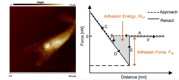
The understanding of adhesive interaction at the nanoscale between functionalized nanoparticles and biological cells is of great importance to develop effective theranostic nanocarriers for targeted cancer therapy. Herein, we use a combination of atomic force microscopy technique and molecular dynamics simulations approach to explore the adhesive interactions at the nanoscale between Triptorelin-conjugated polyethylene glycol (PEG)-coated magnetite nanoparticles and normal/cancerous breast cells. Our findings expand the fundamental understanding of nanoparticle/cell adhesion and provide guidelines for the design of more rational nanocarriers.
Relevant publications: JMBBM (2017), Acta Biomater. (2018)
Cell Viscoelastic Properties
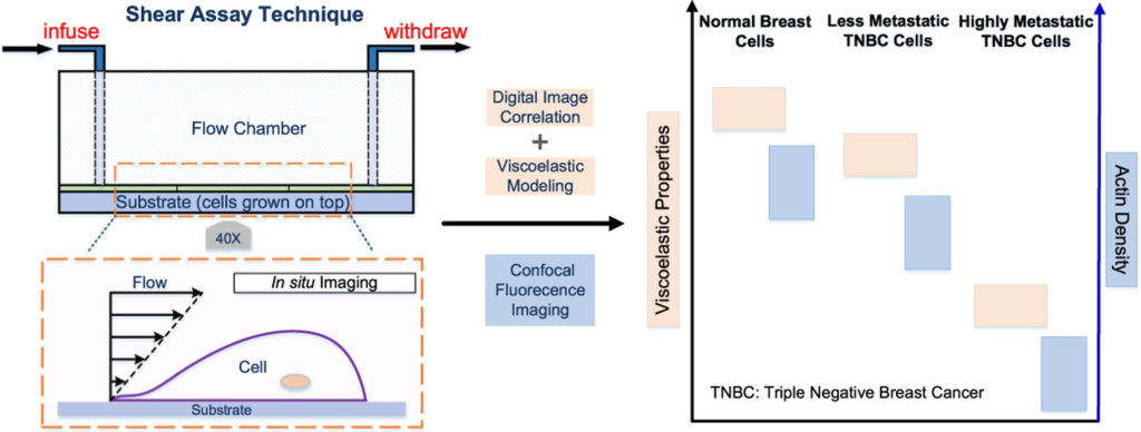
An improved understanding of the evolution of cell structure and viscoelasticity with cancer malignancy could enable the development of a new generation of biomarkers and methods for cancer diagnosis. I studied the viscoelastic properties (moduli and viscosities) and the actin cytoskeletal structures of triple negative breast cancer (TNBC) cells with different metastatic potential. This was achieved through combined shear assay and digital image correlation approach.
Self-assembly
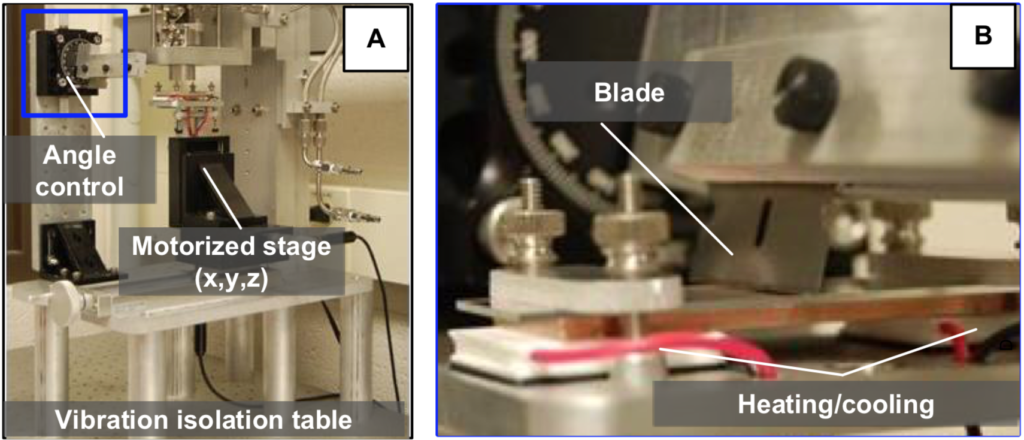
Convective self-assembly offers promise to enable the production of well-ordered films of engineered micro- and nanoparticles at an industrial scale. Using a costumer built blade-casting device, I investigated how blade-casting parameters influence crystal quality, using polystyrene (PS) particles as a model system with optical microscopy and scanning electron microscopy.
Relevant publication: IEEE Nanotechnology (2017)
Quasicrystals
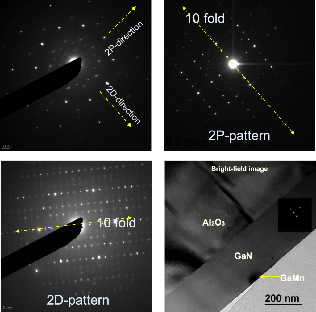
There is a debate in physics and material science arguing that whether quasictrystal phases could also be ferromagnetic. The goal is to use the scanning probe microscope (SPM) that can be performed in magnetic force microscopy (MFM) mode to study the GaMn specimens that have been looked at in transmission electron microscopy (TEM). The goal is to correlate the MFM image features to the TEM image features and figure out which phases are ferromagnetic. Under the supervision of Dr. Kai Sun, I conducted GaMn sample preparation for MFM and TEM, and assisted with material characterization.